

Article Sidebar

 CC BY 4.0
Open access
CC BY 4.0
Open access
You may copy, redistribute and adapt this article, provided that you:
-
Credit the authors and the original article
© 2026 Journal of Tropical Science and Engineering
Integrative Morphological and Molecular Insights into Sacculina lata (Rhizocephala: Sacculinidae) and its Effects on the Host Crab Podophthalmus vigil in Nha Trang Bay
Main Article Content
Abstract
Rhizocephalans are highly specialized parasitic crustaceans commonly infesting decapod hosts, including swimming crabs, and often exert profound effects on host growth and development. Their extremely reduced morphology, however, makes taxonomic identification difficult. In this context, molecular genetic tools have become increasingly valuable for resolving taxonomic relationships within the group. Nevertheless, genetic information in this group remains limited, and only a few representative species have been investigated in detail regarding their biological effects on hosts. For many other rhizocephalans, the extent of host impact remains largely unexplored. The rhizocephalan Sacculina lata has been recorded parasitizing the portunid crabs Charybdis miles and Podophthalmus vigil in the western Pacific region, including Vietnam. Despite these records, the parasite’s morphology has been documented only through schematic illustrations, while detailed photographic documentation, molecular data, and information on host impacts have not been reported. Accordingly, this study employed an integrative morphological and molecular approach to investigate S. lata infesting swimming crabs (Podophthalmus vigil) collected from Nha Trang Bay, Khanh Hoa, Vietnam. Direct observation of fresh specimens, scanning electron microscopy, and histological sections provided a more comprehensive characterization of the parasite’s morphological features. Among 542 examined crabs, infestation prevalence was low (3.51%), showed a clear relationship with host size, and was not associated with host sex. Analyses of nuclear (18S rRNA) and mitochondrial (COI) markers clarified the phylogenetic position of S. lata, revealing a close relationship with Sacculina angulata and several congeners. Parasitism induced pronounced feminization of the abdomen and secondary sexual traits in male hosts, whereas infested females exhibited no significant abdominal modification. Overall, these findings provide new taxonomic, molecular, and ecological insights into rhizocephalan parasitism in an economically important swimming crab species.
Keywords
excrescences, feminization, morphological modifications, Podophthalmus vigil, prevalence, retinacula, Sacculina angulata, Sacculina lata
Article Details
Introduction
Rhizocephala represents a highly specialized group of parasitic barnacles that infest other crustaceans, including swimming crabs of the family Portunidae (Boschma, 1955; Høeg & Lützen, 1995). Rhizocephalans are characterized by complex life cycles and extreme morphological and biological modifications associated with a parasitic mode of life. Infestation is initiated at the cypris larval stage, during which the parasite injects a vermigon into the host’s body. Once inside the host, the parasite develops an internal root-like system (the interna) that spreads throughout host tissues to absorb nutrients, eventually giving rise to an external reproductive structure (the externa) that typically protrudes from the abdominal region of the crab (Glenner et al., 2010; Høeg, 1995; Høeg et al., 2020).
The taxonomy of Rhizocephala has traditionally relied largely on the morphology of the externa (Boschma, 1955; Glenner et al., 2010). However, the extreme structural reduction associated with their highly specialized parasitic lifestyle has resulted in a paucity of reliable diagnostic characters, rendering species delimitation based solely on external features inherently problematic. In response to these limitations, integrative taxonomic approaches combining gross morphology, anatomical dissection, histological analysis, and scanning electron microscopy have been increasingly employed to reveal subtle yet informative characters (Golubinskaya et al., 2021; Lützen et al., 2016; Noever et al., 2016). In parallel, molecular genetic tools have become indispensable for accurate species identification and for resolving phylogenetic relationships within Rhizocephala (Glenner et al., 2003; Glenner & Hebsgaard, 2006; Glenner et al., 2021; Høeg et al., 2020). Nevertheless, despite their recognized value, genetic data remain strikingly limited, with publicly available sequences representing only a small fraction of the approximately 190 described rhizocephalan species (Høeg et al., 2020). Accordingly, expanding integrative morphological and molecular investigations across broader taxonomic and geographic scales is essential for refining species boundaries and achieving a more robust understanding of rhizocephalan diversity and evolution.
Beyond their taxonomic significance, rhizocephalan parasites exert profound effects on their crustacean hosts. Rhizocephalan parasitism induces a wide range of morphological, physiological, and behavioral alterations, frequently culminating in functional or complete parasitic castration (Goddard et al., 2005). Among these effects, modifications of the host reproductive system are particularly conspicuous (Kristensen et al., 2012; Waiho et al., 2017). Gonadal development is often suppressed, while the abdominal region, where the parasite’s externa emerges and may mimic the host’s egg mass, undergoes substantial restructuring. Such changes may involve alterations in both the segmentation pattern and relative size of the abdominal somites. In male hosts, these effects commonly include degenerative transformations accompanied by the acquisition of female-like morphological characteristics (Oanh et al., 2025; Yang et al., 2018).
Within this broader biological and taxonomic context, Sacculina lata Boschma, 1933 (Sacculinidae) has been reported parasitizing two portunid crab hosts, Charybdis miles (De Haan, 1835) and Podophthalmus vigil (Fabricius, 1798), across the western Indo-Pacific region, including Japan, China, Vietnam, and Singapore (Boschma, 1933, 1954; Yang et al., 2018). These host species are widely distributed and of considerable fisheries importance in the region. Although S. lata has been formally described and its diagnostic characters recognized, existing accounts are largely based on schematic line drawings, with a conspicuous absence of photographic documentation capturing the parasite’s morphology in situ and in detail. Moreover, molecular genetic information for S. lata is currently unavailable, and both the infestation status and biological effects of this parasite on P. vigil have received limited attention to date.
To address these knowledge gaps, the present study provides a comprehensive redescription of Sacculina lata based on detailed morphological analyses supported by light microscopy and scanning electron microscopy, together with high-resolution photographic documentation. Molecular sequence data are further integrated to clarify its phylogenetic placement within Rhizocephala. In addition, the infestation status of P. vigil populations is systematically assessed, and parasite-induced morphological alterations in the host are examined. Collectively, these results contribute new insights into the taxonomy, phylogenetic relationships, and ecological implications of rhizocephalan parasitism in swimming crabs.
Materials and Methods
Crab Sampling and Rhizocephalan Collection
A total of 542 specimens of Podophthalmus vigil were collected from commercial fishing vessels operating in Nha Trang Bay, Khanh Hoa Province, Vietnam, between February and July 2025, with approximately 90 individuals sampled per month. In the laboratory, crabs were sorted by sex, and carapace width (CW) was recorded using digital calipers. Each individual was subsequently inspected for rhizocephalan infestation. When present, rhizocephalan parasites were gently removed from the host and preserved for detailed morphological examination and subsequent molecular analyses.
Female crabs possess seven clearly differentiated abdominal somites (segments 1–7), whereas males typically exhibit five externally visible abdominal somites (segments 1, 2, fused 3–5, 6, and 7). To quantitatively assess the effects of rhizocephalan infestation on host abdominal morphology, abdominal width was measured at two standardized positions. In individuals with seven abdominal segments, measurements were taken across the maximum transverse width of segments 6 and 4. In crabs with five abdominal units, measurements were recorded at the widest point of segment 6 and at the second lateral expansion of fused segments 3–5. All measurement landmarks are illustrated in Figure 6. Representative rhizocephalan specimens used for detailed morphological dissection and combined molecular analyses are listed in Table 1.
Morphological Identification
Rhizocephalan parasites were described and identified through detailed examination of the externa, combining gross morphological observations, microanatomical analyses, and scanning electron microscopy (SEM). Gross morphology was assessed using fresh specimens or individuals preserved in 70% ethanol, examined under an Olympus SZ61 stereomicroscope and an Olympus CX41 compound microscope. Information on the specimens examined in detail is provided in Table 1.
Table 1. Measurements and sampling dates of rhizocephalan externae recorded from Podophthalmus vigil in Nha Trang Bay. An asterisk (*) indicates specimens used for molecular analysis
Sample ID | Date collected | Externa dimensions: length × width × height (mm) | Host sex | Host CW (mm) | Intended examination |
1 | 14 Mar 2025 | 25 × 16 × 9* | Male | 48 | Fix and stain, Microanatomy |
2 | 22 Apr 2025 | 8 × 5 × 2 | Female | 53 | Fresh, Gross morphology |
3 | 12 May 2025 | 14 × 7 × 3 | Male | 44 | |
4 | 16 May 2025 | 10 × 6 × 3 | Male | 57.5 | |
5 | 25 Jun 2025 | 20 × 12 × 6* | Female | 64 | |
6 | 25 Jun 2025 | 13 × 8 × 3 | Male | 54 | |
7 | 14 Jul 2025 | 26 × 14 × 8 | Male | 115 | |
8 | 21 × 4 × 8 | ||||
9 | 22 Jul 2025 | 16 × 9 × 4 | Female | 35 | |
10 | 22 Jul 2025 | 21 × 14 × 9 | Female | 58 |
For microanatomical investigation, fresh externae were fixed in 10% neutral buffered formalin, dehydrated through a graded ethanol-xylene series, and embedded in paraplast. Transverse sections approximately 5 μm in thickness were prepared using a Slee 5062 microtome. Sections were stained with hematoxylin and eosin and examined with an Olympus CX41 compound microscope equipped with a Canon R digital camera for photomicrographic documentation.
For SEM analysis of the externa mantle cuticle, specimens were dehydrated through an ascending ethanol series, mounted on aluminum stubs, sputter-coated with gold using an EMS–550 coater, and examined with a FEI Quanta 250 scanning electron microscope. The final composite figures were prepared using PhotoFiltre.
For molecular analyses, a small ventral fragment (approximately 2 mm³) of the Rhizocephalan visceral mass was dissected and preserved in 99.9% ethanol until DNA extraction.
Molecular Analyses
Genomic DNA was isolated from preserved Rhizocephala tissue (specimens subjected to genetic analysis are indicated by an asterisk (*) in Table 1) using the DNeasy Blood & Tissue Kit (Qiagen, Germany) following the manufacturer’s protocol. Partial fragments of the nuclear 18S rDNA and mitochondrial cytochrome c oxidase subunit I (COI) genes were amplified by polymerase chain reaction (PCR) using the primer pairs 329F/aR (Spears et al., 1994) and LCO1490/HCO2198 (Folmer et al., 1994), respectively. Each PCR was performed in a total reaction volume of 25 μL, consisting of 12.5 μL of GoTaq® Green Master Mix (Promega, USA), 1 μL of each primer (10 μM), 3 μL of genomic DNA template, and 7.5 μL of nuclease-free water.
PCR cycling conditions consisted of an initial denaturation at 94 °C for 3 min, followed by 30 cycles of denaturation at 94 °C for 30 s, annealing at 52 °C for 45 s, and extension at 72 °C for 1 min for the 18S rDNA fragment. Amplification of the COI gene employed 35 cycles of 94 °C for 30 s, 42 °C for 45 s, and 72 °C for 30 s. PCR products were electrophoresed on 1% agarose gels and visualized against an appropriate molecular size standard to verify amplification success and fragment length.
Amplified products were purified using a commercial DNA purification kit (Promega) and bidirectionally sequenced with the BigDye Terminator v3.1 Cycle Sequencing Kit (Applied Biosystems), using the same primers as in PCR. Raw sequence reads were assembled and edited using Vector NTI Advance v11.5 (Invitrogen, Life Technologies), and multiple sequence alignments were performed in BioEdit v7.0.5.3 (Hall, 1999). All newly generated sequences were deposited in GenBank.
Sequence similarity was assessed through BLAST searches against the GenBank database. Phylogenetic reconstructions were conducted using maximum-likelihood (ML) and neighbor-joining (NJ) approaches. The best-fitting nucleotide substitution model for ML analysis was selected using ModelTest (Paradis & Schliep, 2019), and the ML tree was inferred in R (Schliep, 2011). NJ analyses were performed in MEGA v7.0 (Kumar et al., 2016). Both phylogenetic analyses incorporated newly obtained 18S rDNA and COI sequences from the present study together with reference sequences retrieved from GenBank (10 sequences for 18S rDNA and 6 for COI mtDNA), using Parasacculina yatsui (Polyascidae) as the outgroup (Table 2).
Table 2. GenBank accession numbers of ingroup and outgroup taxa with corresponding host species included in the phylogenetic analyses
№ | Rhizocephalan species | Host species | Location | Genbank accession number of Rhizocephala | |
COI mtDNA | 18S rDNA | ||||
1 | Heterosaccus lunatus Phillips, 1978 | Charybdis (Charybdis) callianassa (Herbst, 1789) | Australia | DQ059778 | EU082414 |
2 | Heterosaccus papillosus | Charybdis (Charybdis) anisodon | Vietnam | PV631193 | OR644332 |
3 | Loxothylacus panopaei Gissler, 1884 | Loxorhynchus grandis Stimpson, 1857 | USA | KU905819 | AY265364 |
4 | Loxothylacus texanus | Callinectes sapidus | USA | – | L26517 |
5 | Sacculina angulata | Charybdis (Charybdis) hellerii (A. Milne-Edwards, 1867) | Vietnam | PQ776427 | OR644327 |
6 | Sacculina carcini Thompson, 1836 | Carcinus maenas (Linnaeus, 1758) | Sweden | DQ059781 | AY520656 |
7 | Sacculina lata | Podophthalmus vigil | Vietnam | PZ261596 | PZ263541 |
8 | PZ276408 | PZ263542 | |||
9 | Sacculina pugettiae | Scyra ferox (Ohtsuchi & Kawamura, 2019) | Russia | – | PP756299 |
10 | Parasacculina yatsui | Pachygrapsus crassipes | Japan | AB197809 | MG604305 |
Prevalence Analysis
Crabs were considered infested when a rhizocephalan externa was observed on the abdomen. For comparative analyses, P. vigil specimens were grouped by sex (male and female) and assigned to three carapace width categories (≤ 60 mm, 60.5–69.5 mm, and ≥ 70 mm), defined solely for statistical comparison and to maintain balanced sample sizes across groups. Infestation prevalence was calculated as the proportion of infested individuals relative to the total number of examined crabs. Differences in prevalence among host groups were assessed using Chi–square tests.
Effects of Infestation Analysis
The effects of rhizocephalan parasitism on host morphology were evaluated using a combination of morphological observations, morphometric measurements, and statistical analyses. Each specimen was examined both macroscopically and under a stereomicroscope (Olympus SZ61) to document abdominal segmentation patterns, including the number and relative dimensions of abdominal segments, as well as the developmental condition of the pleopods. Abdominal dimensions were quantified by measuring the width of the fourth and sixth abdominal segments (4AW and 6AW).
To explore size-related patterns in abdominal morphology, scatter diagrams were constructed to illustrate the relationship between carapace width (CW) and abdominal segment width. Analyses were conducted separately for four host categories: normal males (NM), infested males (IM), normal females (NF), and infested females (IF). For each category, CW was plotted against both 6AW and 4AW using Microsoft Excel 2016.
Before inferential analyses, data distributions were evaluated for normality using the Shapiro–Wilk test. The effects of infestation status on abdominal dimensions (6AW and 4AW) were assessed using one–way analysis of covariance (ANCOVA), with CW included as a covariate. Pairwise differences among groups were examined using post hoc contrast analyses. All statistical procedures were conducted in SPSS version 20, and statistical significance was set at p < 0.05.
Results
Parasite Taxonomy
Subclass: Cirripedia Burmeister, 1834
Infraclass: Rhizocephala Müller, 1862
Family: Sacculinidae Lilljeborg, 1861
Genus: Sacculina Thompson, 1836
Species: Sacculina lata Boschma, 1933
Collection locality: Nha Trang Bay, Khanh Hoa Province, Vietnam
Host crabs: Podophthalmus vigil (Fabricius, 1798)
Examined samples: 10 individuals (Table 1)
Description
Externa ovoid to trapezoidal (Figure 1C-E). Mantle cuticle thin and densely ornamented with truncated, polygonal pyramid-shaped excrescences (height: 5–8 µm; width: 5–12 µm) (Figure 3A, B). Retinacula scattered, with 4–12 barbed spindles (length: 8–15 µm) arising from a common base (Figure 3C–F).
Visceral mass internally attached to stalk by thin mesentery (Figure 4B, C). Paired receptacles, situated near the lower margin of visceral mass, are cylindrical and mostly straight, with slight anterior curvature at their dorsal ends; they are dorsally separate but ventrally confluent (Figure 4A-D). Vasa deferentia paired and straight, entering receptacles gradually (Figure 4A). Colleteric glands situated anteriorly, consisting of approximately 126 highly branched tubules (Figure 4E) lined with a dense layer of scaly chitin (Figure 4F).
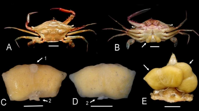
Figure 1. External morphology of Podophthalmus vigil infested by Sacculina lata: A. dorsal view of host crab; B. ventral view of host crab; externa of S. lata visible C. left lateral view of S. lata, D. right lateral view of S. lata, E. double infestation of S. lata. Arrows indicate S. lata; 1. mantle opening; 2. stalk. Scale bars: 5 mm
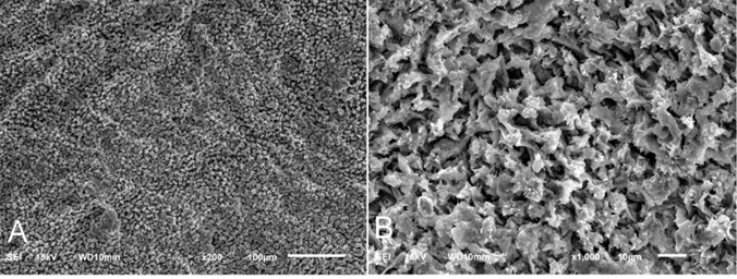
Figure 2. Scanning electron micrograph showing external cuticle of the mantle of Sacculina lata parasitizing on Podophthalmus vigil, densely ornamented with excrescences. A. magnification 200×; B. magnification 1,000×
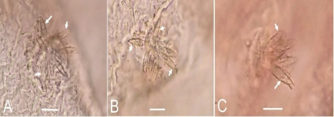
Figure 3. Micrographs of retinacula on the internal mantle cuticle of Sacculina lata parasitizing Podophthalmus vigil, showing 8 (A), 9 (B), and 6 (C) spindles (arrows), respectively. Scale bars: 10 µm
Remarks
Morphological examination shows that the Sacculina lata specimens examined in this study closely conform to the original description and subsequent accounts of the same host species in the same geographic region. Within the genus Sacculina, S. lata exhibits the greatest morphological affinity with Sacculina angulata, a rhizocephalan parasite of Thalamita sima H. Milne Edwards, 1834, Charybdis (Charybdis) orientalis Dana, 1852 and Charybdis (Charybdis) variegata (Fabricius, 1798) (Boschma, 1955; Huang & Lützen, 1998). Nevertheless, the two species can be reliably distinguished by the structure of the external excrescences and receptacles. In S. lata, the excrescences are papilla-shaped, with paired receptacles connected at both extremities and separated medially, whereas in S. angulata the excrescences are truncated and polygonal, with receptacles fused ventrally and separated dorsally.
Molecular validation using BLASTn searches of both nuclear and mitochondrial markers did not identify any reference sequences in GenBank that matches the analyzed material. Sequence similarity to the closest available matches ranged from 95.5% to 99.7% for the nuclear 18S rRNA gene and from 83.7% to 94.2% for the mitochondrial COI gene. These results indicate a clear genetic separation between Sacculina lata and its congeners, at least those for which sequences are available. Consistent with patterns observed in comparative morphology, molecular analyses further suggest a close phylogenetic affinity between S. lata and S. angulata. Pairwise genetic distances between the two species reached 8.1% for the COI gene (Table 3) and 0.3% for the 18S rRNA gene (Table 4). In contrast, no intraspecific sequence divergence was detected among S. lata specimens for either genetic marker, providing strong support for the taxonomic coherence and genetic identity of the species.
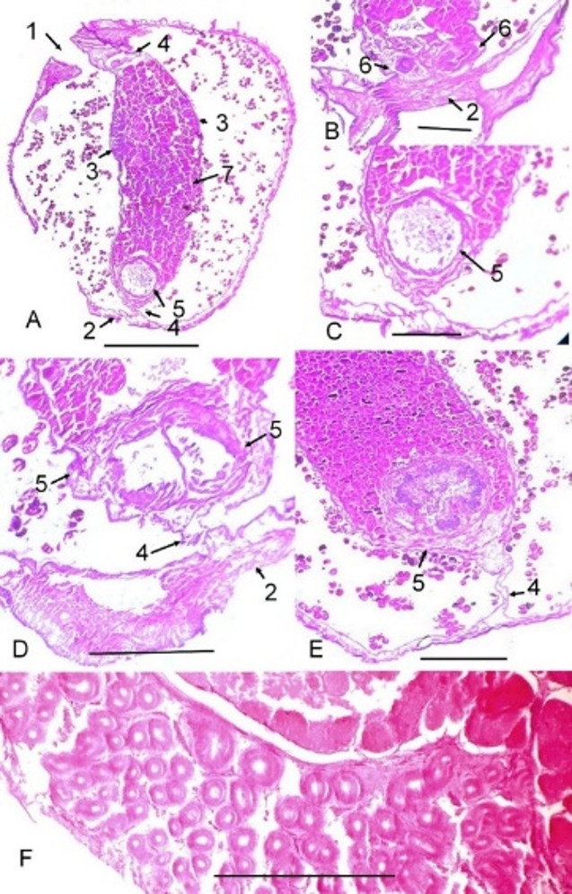
Figure 4. Longitudinal histological sections illustrating internal structures of the externa of Sacculina lata. A. Section passing through the central region of the externa; B. Section through the vas deferens; C. Section at the ventral region where the two receptacles are connected; D. Section at a level where the receptacles are separated; E. Section showing separated receptacles on the dorsal side; F. Colleteric gland. 1. mantle opening; 2. stalk; 3. colleteric glands; 4. mesentery; 5. receptacle; 6. vas deferens; 7. visceral mass. Scale bars: A = 1.0 mm; B-F = 0.5 mm
Phylogeny
Phylogenetic relationships among selected Rhizocephala species were reconstructed using maximum likelihood (ML) and neighbor-joining (NJ) approaches based on mitochondrial and nuclear gene datasets. The COI-based phylogeny (Figure 5A) consistently
recovered the genus Sacculina as being more closely related to Heterosaccus than to Loxothylacus. Within Sacculina, S. lata and S. angulata formed a strongly supported sister clade, clearly separated from other congeners with a high bootstrap value (100%), indicating robust mitochondrial divergence.
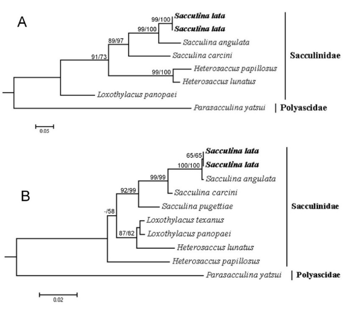
Figure 5. Inferred phylogenetic position of Sacculina lata (in bold) among representative Rhizocephala taxa from maximum-likelihood and neighbor–joining analyses. Trees were reconstructed from (A) COI mitochondrial sequences under the TVM+I+G substitution model and (B) 18S rRNA nuclear sequences under the GTR+G(4)+I model. Bootstrap support (ML/NJ; 1,000 pseudoreplicates) is indicated only for nodes receiving ≥ 50% support
In contrast, the 18S rRNA tree (Figure 5B) revealed a clear genetic separation between Sacculina and the remaining genera within Sacculinidae. In this topology, S. lata clustered very closely with S. angulata, suggesting limited species–level resolution when inferred from the conserved nuclear marker alone. Nevertheless, the distinct separation observed in the COI phylogeny supports their recognition as separate species. Overall, integrating mitochondrial and nuclear gene datasets yields a more complete and robust phylogenetic reconstruction of Rhizocephala than either marker analyzed independently.
Table 3. Genetic divergence based on COI p-distances (%) among analyzed rhizocephalan species
| Rhizocephala species | 1 | 2 | 3 | 4 | 5 | 6 | 7 | 8 |
1 | Heterosaccus papillosus | ID | 21.6 | 23.0 | 22.6 | 22.6 | 6.1 | 24.6 | 30.8 |
2 | Sacculina carcini |
| ID | 17.5 | 16.1 | 6.1 | 21.3 | 22.1 | 31.9 |
3 | Sacculina angulata |
|
| ID | 8.1 | 8.1 | 21.9 | 22.6 | 30.7 |
4 | Sacculina lata |
|
|
| ID | 0.0 | 20.8 | 21.4 | 30.3 |
5 | Sacculina lata |
|
|
|
| ID | 20.8 | 21.4 | 30.3 |
6 | Heterosaccus lunatus |
|
|
|
|
| ID | 24.1 | 30.2 |
7 | Loxothylacus panopaei |
|
|
|
|
|
| ID | 29.2 |
8 | Parasacculina yatsui |
|
|
|
|
|
|
| ID |
Table 4. Genetic divergence based on 18S p–distances (%) among analyzed rhizocephalan species
| Rhizocephalan species | 1 | 2 | 3 | 4 | 5 | 6 | 7 | 8 | 9 | 10 |
1 | Sacculina lata | ID | 0.0 | 0.3 | 2.3 | 5.0 | 7.3 | 6.7 | 5.9 | 5.8 | 13.6 |
2 | Sacculina lata |
| ID | 0.3 | 2.3 | 5.0 | 7.3 | 6.7 | 5.9 | 5.8 | 13.6 |
3 | Sacculina angulata |
|
| ID | 2.2 | 4.9 | 7.2 | 6.6 | 5.8 | 5.7 | 13.6 |
4 | Sacculina carcini |
|
|
| ID | 4.0 | 6.6 | 5.6 | 5.0 | 4.9 | 12.8 |
5 | Sacculina pugettiae |
|
|
|
| ID | 5.9 | 5.1 | 4.1 | 4.1 | 12.6 |
6 | Heterosaccus papillosus |
|
|
|
|
| ID | 5.7 | 5.0 | 4.8 | 12.5 |
7 | Heterosaccus lunatus |
|
|
|
|
|
| ID | 2.5 | 2.1 | 11.5 |
8 | Loxothylacus texanus |
|
|
|
|
|
|
| ID | 0.7 | 11.4 |
9 | Loxothylacus panopaei |
|
|
|
|
|
|
|
| ID | 11.4 |
10 | Parasacculina yatsui |
|
|
|
|
|
|
|
|
| ID |
Prevalence of Infestation
A total of 542 specimens of Podophthalmus vigil were examined, comprising 355 males and 187 females. Among these, 19 individuals (12 males and 7 females) were infested with Sacculina lata, resulting in an overall prevalence of 3.51% (Table 5). Single parasite infections predominated, except for one male crab that harbored two rhizocephalans. Although the prevalence in females (3.74%) was slightly higher than that in males (3.38%), this difference was not statistically significant (χ² = 0.0477, df = 1, p > 0.05). In contrast, infestation prevalence differed significantly among the three crab size classes (χ² = 12.40, df = 2, p < 0.01). Specifically, the smallest size class exhibited the highest prevalence (7.26%), followed by the intermediate size class (60.5–69.5 mm; 2.72%. In contrast, only a single infested individual was recorded in the largest size class (≥ 70 mm), corresponding to a prevalence of 0.56% (Table 5). Overall, these results indicate that infestation by S. latain P. vigil is more strongly associated with host size than with host sex.
Table 5. Overall, sex–specific, and size–class–specific prevalence of Sacculina lata infesting Podophthalmus vigil. Different lowercase letters (a, b) indicate statistically significant differences in infestation prevalence among crab size classes (p < 0.05)
Overall prevalence (%) | Prevalence by sex (%) | Prevalence by size categories (%) | |||
Male | Female | ≤ 60 mm | 60.5–69.5 mm | ≥ 70 mm | |
n = 542 | n = 355 | n = 187 | n = 179 | n = 184 | n = 179 |
3.51 | 3.38 | 3.74 | 7.26a | 2.72b | 0.56b |
Effects of Infestation
In comparison of the abdominal morphology of uninfested Podophthalmus vigil (Figure 6A-D) with that of individuals infested by Sacculina lata, marked morphological alterations were documented. In all infested males (n = 12), pronounced feminization was observed, characterized by transformation of the abdomen into a distinctly seven–segmented form, with well–developed setae along the lateral margins of each segment, resembling the typical female condition. Nevertheless, the pleopods retained the diagnostic male morphology and did not exhibit branching or reduction (Figure 6E, F). In contrast, infested females (n = 7) showed no apparent morphological modifications, with both the number of abdominal segments and pleopod structure remaining comparable to those of uninfested females (Figure 6G, H). Overall, these observations indicate that S. lata infestation induces substantial, sex-specific morphological modifications of the abdomen in P. vigil, predominantly affecting male hosts.
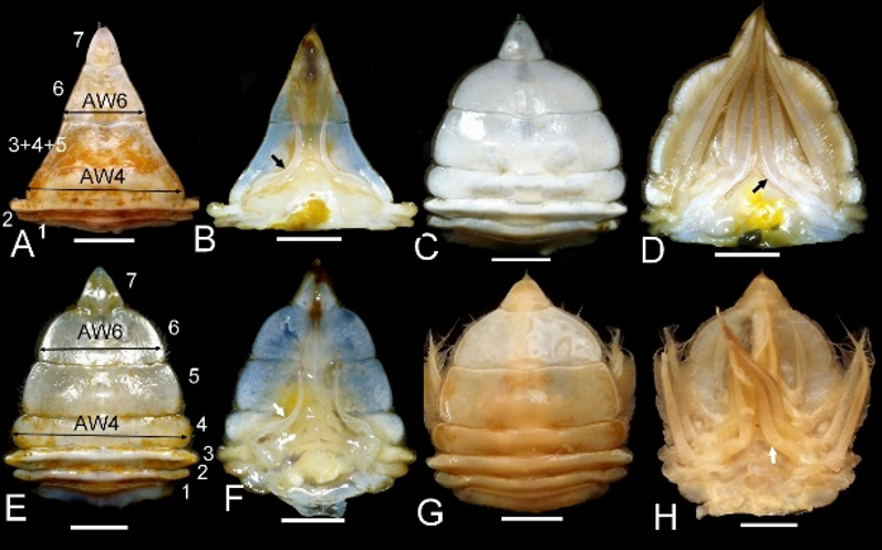
Figure 6. Morphological modifications of the abdomen of the crab Podophthalmus vigil induced by infestation by the rhizocephalan Sacculina lata. A–B. Dorsal and ventral views of the abdomen of a normal male. C–D. Dorsal and ventral views of the abdomen of a normal female. E–F. Dorsal and ventral views of the abdomen of an infested male. G–H. Dorsal and ventral views of the abdomen of a female infested with S. lata. Arrows indicate pleopods. Scale bars: 5 mm
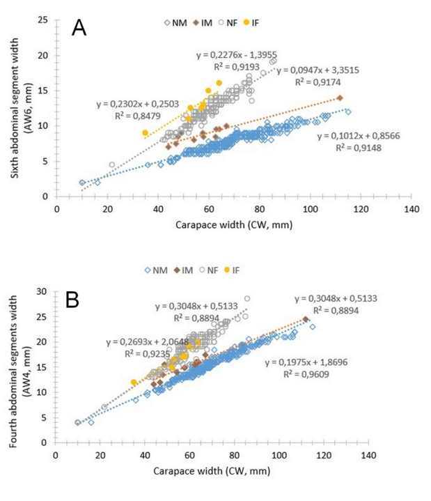
Figure 7. Relationship between carapace width (CW) and abdominal segment dimensions in Podophthalmus vigil. Scatterplots illustrate the association of CW with width of the sixth abdominal segment (6AW) (A) and width of the fourth abdominal segment (4AW) (B) in crabs infested and uninfested with Sacculina lata. IM. infested male; NM. normal male; IF. infested female; NF. normal female
Scatterplot analyses revealed a strong positive relationship between carapace width and the widths of both the sixth and fourth abdominal segments across all examined groups, including uninfested males, uninfested females, infested males, and infested females (r > 0; R² > 0.84; Figure 7A, B). This consistent scaling relationship indicates that abdominal segment width generally increases with overall body size regardless of infestation status or sex. After accounting for the effect of carapace width, analysis of covariance (ANCOVA) detected significant differences in the adjusted mean width of the sixth abdominal segment between infested and uninfested individuals of both sexes, with marked effects observed in males (F = 280.457, df = 1, p < 0.01) as well as in females (F = 48.157, df = 1, p < 0.01). These results demonstrate that infestation by Sacculina lata is associated with an enlargement of the sixth abdominal segment in both male and female crabs.
In contrast, the response of the fourth abdominal segment exhibited a clear sex-specific pattern. A statistically significant difference in its width was detected only between infested and uninfested males (F = 103.320, df = 1, p < 0.01). In contrast, no significant difference was observed between infested and uninfested females (F = 1.672, df = 1, p > 0.05). Overall, these findings indicate that while S. lata infestation affects abdominal morphology in both sexes, its impact is more pronounced and consistently expressed in male than in female hosts.
Discussion
Rhizocephalans represent one of the most extreme examples of morphological and biological specialization among parasitic crustaceans. Their highly reduced body plan, coupled with the loss of most conventional diagnostic characters, has long posed challenges for species delimitation and comparative taxonomy. Sacculina lata has previously been reported from two portunid hosts, Charybdis miles and Podophthalmus vigil, distributed along the western Pacific coast (Boschma, 1933, 1954). However, detailed morphological documentation of this species has remained limited. In the present study, we adopted an integrative morphological approach, enabling a more comprehensive characterization of the parasite’s structural organization and revealing several diagnostic features that have not been clearly illustrated before.
In particular, the excrescences on the external cuticle of the mantle were documented in detail and shown to possess distinct structural characteristics. These excrescences differ markedly from those described for Sacculina angulata, which has been regarded as the most morphologically similar congener (Boschma, 1955). The clear differentiation observed in these mantle structures provides new morphological evidence supporting the separation of the two species, complementing earlier taxonomic assessments that relied largely on line drawings and limited external descriptions. Consequently, the present study contributes novel, image-based morphological data that substantially refine the diagnosis of S. lata.
From a molecular perspective, phylogenetic analyses of both nuclear 18S rRNA and mitochondrial COI genes revealed patterns largely congruent with morphological observations. Both markers consistently placed S. lata and S. angulata as closely related taxa, supporting their affinity within Sacculinidae. Notably, species-level separation was strongly supported by the COI phylogeny, in which the two taxa formed well-resolved, distinct clades with maximal bootstrap support (100%). In contrast, the 18S rRNA fragment (approximately 1,100 bp) exhibited very low genetic divergence between the two species (0.3%) (Table 4), reflecting the conservative nature of nuclear ribosomal genes. These results highlight the effectiveness of COI as a barcode marker for rhizocephalan taxonomy, while suggesting that nuclear markers may capture deeper evolutionary signals and require longer sequences or multiple primer sets to provide sufficient phylogenetic resolution. The combined use of mitochondrial and nuclear data therefore remains essential for robust species delimitation and evolutionary inference in this group.
Although molecular data for Sacculina lata are presented here for the first time, its infestation dynamics have not been entirely unexplored. A previous study provided a detailed and systematic assessment of rhizocephalan infestation in Charybdis miles from the Gulf of Tonkin, Vietnam (Yang et al., 2018). This work constitutes a rigorous baseline for comparison, enabling a more informed evaluation of infestation patterns observed in the present study. In comparison with that earlier investigation, the prevalence of S. lata infesting Podophthalmus vigil recorded here was relatively low (3.51%) and showed no significant difference between male and female hosts. In contrast, the previous study on C. miles reported a higher overall prevalence (7.4%), with female crabs exhibiting a significantly higher infestation rate than males. These discrepancies may reflect differences in geographic context and host–specific ecological traits, all of which may influence exposure risk and host-parasite interactions.
The study by Yang et al. (2018) did not explicitly analyze infestation prevalence across host size classes; however, it demonstrated that infested crabs had a significantly smaller mean body size than uninfested individuals. This pattern is consistent with the well-established biological effects of rhizocephalans, which suppress molting and interfere with somatic growth, thereby preventing infested hosts from attaining larger sizes (Høeg, 1995). Although changes in abdominal segmentation were not addressed in the earlier study, male feminization was clearly documented, as evidenced by an enlarged abdomen relative to uninfested males. This modification has been interpreted as a parasite-induced alteration that enhances protection and functional accommodation of the externa.
When the host impacts of S. lata are compared with those induced by Heterosaccus papillosus infesting Charybdis (C.) anisodon in the same biogeographic region, notable differences emerge. While H. papillosus induces pronounced feminization in both male and female hosts, often resulting in the development of pleopods in males and obscuring sexual dimorphism (Oanh et al., 2025), S. lata appears to exert disproportionately stronger effects on male hosts, with more limited morphological modification in females. Importantly, these rhizocephalans belong to two different genera, suggesting that the observed differences in host manipulation may reflect species-specific traits rather than a shared strategy at the generic or familial level. Consequently, further comparative studies encompassing multiple rhizocephalan species and host taxa are required to determine whether such host effects represent adaptive strategies at the species level or indicative of broader evolutionary patterns at higher taxonomic ranks.
Conclusion
In conclusion, the present study provides the first integrative morphological and molecular characterization of Sacculina lata infesting Podophthalmus vigil. By integrating fresh observations, ultrastructural and histological analyses, and mitochondrial and nuclear genetic data, diagnostic features that distinguish S. lata from closely related congeners are clarified, and its phylogenetic position within Sacculinidae is confirmed. Moreover, the assessment of infestation prevalence and host impacts reveals host- and sex-specific patterns that differ from those reported in previous studies on other portunid crabs, highlighting variability in host-parasite interactions among rhizocephalans. Collectively, these findings not only refine the taxonomy of S. lata but also contribute new insights into the diversity of parasitic strategies and their ecological consequences in brachyuran hosts.
Funding: This research was financially supported by the Joint Vietnam-Russia Tropical Science and Technology Research Center under Branch-level Project VBĐ2.02/25.
Statement on the use of Generative AI: The authors declare that AI tools were used only for language editing/formatting, and not for generating scientific content. All data, analyses, and interpretations were performed and verified by the authors, who take full responsibility for the manuscript.
Author contributions: Le Thi Kieu Oanh: Conceptualization, Formal Analysis, Investigation, Data Curation; Writing - Original Draft, Writing - Review & Editing, Project Administration; Vo Thi Ha, Nguyen Phuong Lien, Hoang Thi Ngoc Anh, and Nguyen Thi Hai Thanh: Investigation; Nguyen Trinh Duc Hieu: Visualization.
Conflicts of interest statement: The authors declare no conflicts of interest.
Data Availability Statement: The data supporting the findings of this study are available from the corresponding author upon reasonable request.
References
Boschma, H. (1933). New species of Sacculinidae in the collection of the United States National Museum. Tijdschrift der Nederlandsche Dierkundige Vereeniging, 3, 219-241.
Boschma, H. (1954). Rhizocephala from Indo-China. Proceedings of the Koninklijke Nederlandse Akademie van Wetenschappen, Series C, 57, 230-241.
Boschma, H. (1955). The described species of the family Sacculinidae. Zoologische Verhandelingen, 27(1), 1-76.
Folmer, O., Black, M., Hoeh, W., Lutz, R., & Vrijenhoek, R. (1994). DNA primers for amplification of mitochondrial cytochrome c oxidase subunit I from diverse metazoan invertebrates. Molecular Marine Biology and Biotechnology, 3(5), 294-299.
Glenner, H., & Hebsgaard, M. B. (2006). Phylogeny and evolution of life history strategies of the parasitic barnacles (Crustacea, Cirripedia, Rhizocephala). Molecular Phylogenetics and Evolution, 41(3), 528-538. https://doi.org/10.1016/j.ympev.2006.06.004
Glenner, H., Høeg, J. T., Rees, D. J., & Schubart, C. D. (2021). Phylogenetic analyses suggest a single origin of freshwater barnacles. In T. Kawai & D. C. Rogers (Eds.), Recent Advances in Freshwater Crustacean Biodiversity and Conservation (pp. 3-18). CRC Press. https://doi.org/10.1201/9781003139560-2
Glenner, H., Høeg, J. T., Stenderup, J., & Rybakov, A. V. (2010). The monophyletic origin of a remarkable sexual system in akentrogonid rhizocephalan parasites: a molecular and larval structural study. Experimental parasitology, 125(1), 3-12. https://doi.org/10.1016/j.exppara.2009.09.019
Glenner, H., Lützen, J., & Takahashi, T. (2003). Molecular and morphological evidence for a monophyletic clade of asexually reproducing Rhizocephala: Polyascus, new genus (Cirripedia). Journal of Crustacean Biology, 23(3), 548-557. https://doi.org/10.1651/C-2361
Goddard, J. H. R., Torchin, M. E., Kuris, A. M., & Lafferty, K. D. (2005). Host specificity of Sacculina carcini, a potential biological control agent of the introduced European green crab Carcinus Maenas in California. Biological Invasions, 7(6), 895-912. https://doi.org/10.1007/s10530-003-2981-0
Golubinskaya, D. D., Korn, O. M., Sharina, S. N., & Selin, N. I. (2021). Sympatric two-species infestation by rhizocephalan barnacle parasites in the spider crab Pugettia aff. ferox Ohtsuchi & Kawamura, 2019 from Peter the Great Bay (Northwestern Sea of Japan). Zoological Studies, 60, e54. https://doi.org/10.6620/ZS.2021.60-54
Hall, T. A. (1999). BioEdit: A user-friendly biological sequence alignment editor and analysis program for Windows 95/98/NT. Nucleic Acids Symposium Series, 41, 95-98.
Høeg, J. T. (1995). The biology and life cycle of the Rhizocephala (Cirripedia). Journal of the Marine Biological Association of the United Kingdom, 75(3), 517-550. https://doi.org/10.1017/S0025315400038996
Høeg, J. T., & Lützen, J. (1995). Life cycle and reproduction in the cirripedia Rhizocephala. Oceanography and Marine Biology: an Annual Review, 33, 427-485.
Høeg, J. T., Noever, C., Rees, D. A., Crandall, K. A., & Glenner, H. (2020). A new molecular phylogeny-based taxonomy of parasitic barnacles (Crustacea: Cirripedia: Rhizocephala). Zoological Journal of the Linnean Society, 190(2), 632-653. https://doi.org/10.1093/zoolinnean/zlz140
Huang, J. F., & Lützen, J. (1998). Rhizocephalans (Crustacea: Cirripedia) from Taiwan. Journal of Natural History, 32(9), 1319-1337. https://doi.org/10.1080/00222939800770661
Kristensen, T., Nielsen, A. I., Jørgensen, A. I., Mouritsen, K. N., Glenner, H., Christensen, J. T., Lützen, J., & Høeg, J. T. (2012). The selective advantage of host feminization: A case study of the green crab Carcinus maenas and the parasitic barnacle Sacculina carcini. Marine Biology, 159(9), 2015-2023. https://doi.org/10.1007/s00227-012-1988-4
Kumar, S., Stecher, G., & Tamura, K. (2016). MEGA7: Molecular Evolutionary Genetics Analysis Version 7.0 for Bigger Datasets. Molecular Biology and Evolution, 33(7), 1870-1874. https://doi.org/10.1093/molbev/msw054
Lützen, J., Itani, G., Jespersen, Å., Hong, J. S., Rees, D., & Glenner, H. (2016). On a new species of parasitic barnacle (Crustacea: Rhizocephala), Sacculina shiinoi sp. nov., parasitizing japanese mud shrimps Upogebia spp. (Decapoda: Thalassinidea: Upogebiidae), including a description of a novel morphological structure in the Rhizocephala. Zoological Science, 33(2), 204-212. https://doi.org/10.2108/zs150112
Noever, C., Olson, A., & Glenner, H. (2016). Two new cryptic and sympatric species of the king crab parasite Briarosaccus (Cirripedia: Rhizocephala) in the North Pacific. Zoological Journal of the Linnean Society, 176(1), 3-14. https://doi.org/10.1111/zoj.12304
Oanh, L. T. K., Hoang, A. T. N., Vu, T. Q., Ha, V. T., Tran, S. Q., Truong, O. T., Glenner, H., & Dang, B. T. (2025). Morphological and molecular characterization of Heterosaccus papillosus Boschma, 1933 (Rhizocephala: Sacculinidae) and its effects on the swimming crab Charybdis anisodon De Haan, 1850 (Decapoda: Portunidae) in Nha Trang Bay, Vietnam. Zootaxa, 5673(2), 213-226. https://doi.org/10.11646/zootaxa.5673.2.3
Paradis, E., & Schliep, K. (2019). Ape 5.0: an environment for modern phylogenetics and evolutionary analyses in R. Bioinformatics (Oxford, England), 35(3), 526-528. https://doi.org/10.1093/bioinformatics/bty633
Schliep K. P. (2011). Phangorn: phylogenetic analysis in R. Bioinformatics (Oxford, England), 27(4), 592-593. https://doi.org/10.1093/bioinformatics/btq706
Spears, T., Abele, L. G., & Applegate, M. A. (1994). Phylogenetic study of cirripedes and selected relatives (Thecostraca) based on 18S rDNA sequence analysis. Journal of Crustacean Biology, 14(4), 641-656. https://doi.org/10.2307/1548858
Waiho, K., Fazhan, H., Glenner, H., & Ikhwanuddin, M. (2017). Infestation of parasitic rhizocephalan barnacles Sacculina beauforti (Cirripedia, Rhizocephala) in edible mud crab, Scylla olivacea. PeerJ, 5, e3419. https://doi.org/10.7717/peerj.3419
Yang, C.-P., Sun, D.-R., Qiu, Y.-S., Liu, Y., Shan, B., Li, T., & Liu, S. (2018). The parasitic barnacle Sacculina lata Boschma, 1933 (Cirripedia, Rhizocephala, Sacculinidae) infecting the commercial swimming crab Charybdis miles (De Haan, 1835) (Decapoda, Portunidae) in the Beibu Gulf, South China Sea. Crustaceana, 91(5), 577-590. https://doi.org/10.1163/15685403-00003787