

Article Sidebar

 CC BY 4.0
Open access
CC BY 4.0
Open access
You may copy, redistribute and adapt this article, provided that you:
-
Credit the authors and the original article
© 2026 Journal of Tropical Science and Engineering
Molecular Characterization and Phylogenetic Relationships of a Novel Pipistrellus ceylonicus-derived Picobirnavirus
Main Article Content
Abstract
The Picobirnaviridae family comprises small, non-enveloped viruses with a bi-segmented, double-stranded RNA genome, exhibiting broad host range and high genetic diversity. Despite over 7,000 entries in public databases, only 53 originate from bats, and merely one contains a near-complete capsid protein sequence, highlighting a significant knowledge gap. This study reports the detection and genomic characterization of a novel picobirnavirus derived from Pipistrellus ceylonicus bat from the Kon Chu Rang Nature Reserve, Gia Lai province, Vietnam. Using metagenomic sequencing, we obtained genomic sequences for both segments (dsRNA1: 1,902 bp; dsRNA2: 1,626 bp), encoding the capsid protein and RNA-dependent RNA polymerase (RdRp), respectively. Phylogenetic analysis of RdRp amino acid sequences robustly placed the virus within the International Committee on Taxonomy of Viruses (ICTV)-recognized genogroup 2 and the proposed genus Alphapicobirnavirus. The obtained capsid protein showed the highest sequence similarity to picobirnaviruses isolated from a human and a macaque (genetic distances of 0.59 and 0.62), while exhibiting a distant relationship (divergence of 0.79) to the only known near-complete bat picobirnavirus capsid sequence. This significant genetic distance, particularly from the sole chiropteran reference, supports the identification of a novel viral species within the Picobirnaviridae family. The characterized virus expands the known diversity of bat-related picobirnaviruses and provides crucial genomic data, contributing to a more accurate understanding of the family's evolution and host range.
Keywords
Bats, Metagenomics, Phylogeny, RNA Viruses, Vietnam, Viral Genomics, Virus Classification
Article Details
Introduction
The Picobirnaviridae family comprises small, non-enveloped viruses with a double-stranded RNA (dsRNA) genome of 4.1–4.6 kbp, consisting of two segments (the larger 2.4–2.7 kbp and the smaller 1.7–1.9 kbp). However, a rare single, fused segment has also been documented (Delmas et al., 2019). Segment 1 contains two or three open reading frames (ORFs). The function of the first ORF, which is non-obligatory, remains unknown, as does that of the second ORF-encoded peptide. The third ORF encodes the precursor of the nucleocapsid protein. Segment 2 contains a single ORF encoding the RNA-dependent RNA polymerase (RdRP).
A single genus, Orthopicobirnavirus, is currently recognized within the family. This genus is subdivided into five distinct genogroups, which exhibit substantial genetic diversity among themselves (Giannitti et al., 2015). Genogroups I and II are predominantly associated with vertebrate hosts, while genogroup III is primarily linked to invertebrates (Shi et al., 2016). Limited information is currently available for the proposed genogroups IV and V (Knox et al., 2018). A proposed alternative taxonomic classification subdivides the family into 7 genera based on RdRP amino acid sequence similarity (Sadiq et al., 2024).
Genomic sequences related to Picobirnaviridae have been identified in a wide range of vertebrates, including mammals, fish, birds (such as chickens and rheas), and humans. Invertebrate-associated picobirnaviruses, in contrast, were discovered through extensive screening of invertebrate transcriptomic datasets (Shi et al., 2016).
Among known picobirnaviruses, those associated with mammals, including humans, are the most extensively characterized group. Potential non-human mammalian hosts include horses (Giannitti et al., 2015), pigs (Ganesh et al., 2012), deer (Huaman et al., 2021), rodents (including marmots) (Peng et al., 2025), bats (Yan et al., 2019) and others. While these viruses are most frequently detected in fecal samples, they have also been identified in the sputum of patients with acute respiratory disease (Berg et al., 2021). There is some evidence linking picobirnaviruses to gastrointestinal illness in humans, particularly among immunocompromised individuals (Giordano et al., 1998; Legoff et al., 2017). However, a definitive causal relationship has never been established, supporting the hypothesis that they are opportunistic agents (Malik et al., 2014). This pattern extends to other mammals as well, with picobirnaviruses detected in both symptomatic and clinically healthy animals.
The inability to cultivate picobirnaviruses in animal cell culture, combined with the lack of a clear virus-disease relationship, raises fundamental questions about their true host range (Kashnikov et al., 2023). Several genetic features of picobirnaviruses suggest they might infect bacteria or fungi rather than animal cells. Supporting evidence includes the presence of Shine-Dalgarno sequences upstream of open reading frames as a hallmark commonly found in bacteriophage genomes (Krishnamurthy & Wang, 2018). Furthermore, some picobirnaviruses use a non-standard genetic code, such as the fungal/invertebrate mitochondrial code (Yinda et al., 2018), and the vast majority of detections occur in bacteria-rich environments, such as the gut. Nevertheless, the definitive host of Picobirnaviridae remains unresolved. Confirmation will likely require either the development of a functional cultivation system complicated by the fact that many gut microbes thought to be the actual hosts are themselves unculturable (Kim et al., 2011), or further structural and functional studies of the viral capsid to identify potential bacterium-specific entry mechanisms.
Chiroptera are recognized as a major reservoir for diverse viruses, and due to their capacity for long-distance movement and occupation of varied biomes, their virome diversity is considerable, even within a single country (Lapshina et al., 2025). While numerous studies have identified diverse viruses in Vietnamese bats (Arai et al., 2019; Inagaki et al., 2020; Latinne et al., 2023), picobirnaviruses had remained undetected before this research. Of the over 7,000 Picobirnaviridae records currently available in public genomic databases, however, only 53 originate from bats, and only one containing a capsid protein sequence longer than 400 amino acids. In this study, we report the detection of a potentially novel picobirnavirus derived from a Pipistrellus ceylonicus bat captured in the Kon Chu Rang Nature Reserve, Gia Lai province, Vietnam. We present the genomic sequences for both viral segments and the corresponding near-complete amino acid sequences of the RNA-dependent RNA polymerase (RdRP) and capsid protein. These sequences, identified through metagenomic sequencing, were used for subsequent molecular and phylogenetic analysis.
Materials And Methods
Sample Collection and Animal Handling
A subadult female P. ceylonicus was captured on June 3, 2025, in the Kon Chu Rang Nature Reserve, Gia Lai province, Vietnam, using a mist net. Taxonomic identification was conducted following standard taxonomic guidelines (Kruskop, 2013). All animal handling procedures were performed by trained personnel in compliance with ethical standards for wildlife research, under a protocol approved by the local ethics committee of the Joint Vietnamese-Russian Scientific Research and Technology Center (Protocol No. 1824 dated 21.05.2025- approval number 1837/CN-HDDDDV dated 21.05.2025). The fecal collection was performed by placing the bat in a sterile bag and waiting for natural excretion. The sample was harvested from the bag using sterile instruments, omitting the surface layer of the produced biosample. Immediately after collection, the fecal sample was placed in a 2.0 ml cryovial, flash-frozen in liquid nitrogen in the field and maintained under these conditions until transfer to the laboratory, where it was stored at -80 °C before the study.
Nucleic Acids Extraction
The fecal sample was homogenized by vortexing until a uniform suspension was achieved. The homogenate was then centrifuged at 17,761g, and the resulting supernatant was subjected to a second round of centrifugation to remove any remaining particulate debris. Total nucleic acids were extracted from 100 µl of the clarified supernatant using the RIBO-prep kit (AmpliTest, Russia) according to the manufacturer's instructions.
Library Preparation and Sequencing
The nucleic acid extract was treated with DNAse I (NEB) and the remaining RNA was purified using MagPure A4 XP magnetic beads (Magen, China) with 1.2:1 beads:sample ratio. The first-strand cDNA synthesis was performed using NEBNext Ultra II RNA First Strand Synthesis Module (New England Biolabs, USA) with denaturation and random priming at 65 °C for 5 minutes. The second strand cDNA synthesis was performed with NEBNext Ultra II RNA Second Strand Synthesis Module (New England Biolabs, USA); the end repair was performed using NEBNext Ultra II End Repair/dA-Tailing Module. The adapter from the PCR Barcoding Expansion 1-96 (Oxford Nanopore Technologies, UK) was ligated using the NEBNext Ultra II Ligation Module (New England Biolabs, USA); the sample was then purified with MagPure A4 beads with 0.3:1 beads:sample volume ratio. Barcoding PCR amplification was performed using PCR barcode primers from PCR Barcoding Expansion 1-96 (Oxford Nanopore Technologies, UK) and BioMaster LR HS-PCR 2x mix (Biolabmix, Russia) with a 5-minutes elongation time. The final third-generation sequencing library was prepared using the Ligation Sequencing Kit V14 (Oxford Nanopore Technologies, UK) and sequenced on the R10.4.1 MINIon flow cell (Oxford Nanopore Technologies, UK) with Nanoporus sequencer (Nanoporus, Russia).
Sequencing Data Analysis and Viral Genome Assembling
Basecalling was performed using Dorado v.7.2.13 in super-accurate mode, yielding approximately 2.25 million reads (1.07 gigabases) for the sample. Adapter trimming and chimera resolution were conducted with Porechop v.0.2.4 (Wick & Volkening, 2017). Reads were assembled de novo into contigs using Canu v.2.2. (Koren et al., 2017), followed by removal of non-target sequences with Kraken2 v2.1.3 (Wood et al., 2019). Taxonomic classification was optimized using a combined nucleotide and protein approach with BLASTn v2.16.0+ (Altschul et al., 1990) and DIAMOND v2.1.16 (Buchfink et al., 2021) to identify Picobirnaviridae contigs. Raw reads were then mapped back to the identified contigs, and the assemblies were polished with Pilon v1.24 (Walker et al., 2014) to correct insertion, deletion, and mismatch errors.
Genome Annotation and Phylogenetic Analysis
Genome annotation, protein translation, identification, and visualization were performed manually using a combination of SnapGene Viewer (Tello et al., 2022) and the protein BLAST (pBLAST) tool (McGinnis & Madden, 2004). A maximum-likelihood phylogenetic tree was constructed from an alignment of RNA-dependent RNA polymerase (RdRP) protein sequences from viruses belonging to the family Picobirnaviridae, using an approach similar to that used in other Picobirnaviridae taxonomy studies (Sadiq et al., 2024). The analysis was performed using RAxML-NG version 1.2.2 (Kozlov et al., 2019) under the LG+I+G substitution model with 1000 bootstrap replicates. Reference sequences included formally classified taxa from the ICTV Picobirnaviridae report as well as proposed genera suggested by recent taxonomic evaluations (Delmas et al., 2019).
Results
A subadult female of P. ceylonicus species was captured on June 3, 2025, in the tropical forest of Kon Chu Rang Nature Reserve, Gia Lai province, Vietnam (Figure 1).
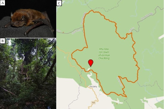
Figure 1. (A) P. ceylonicus captured in the Kon Chu Rang Nature Reserve, Gia Lai province, Vietnam (Photo: Yuzefovich A.P.). (B) Undisturbed forest habitat in Kon Chu Rang Nature Reserve, Gia Lai province, Vietnam, where the P. ceylonicus was captured (Photo: Yuzefovich A.P.). (C) Geographic location of the capture site (14.474; 108.542)
Genome sequencing and assembly yielded sequences for two segments: dsRNA1 (1,902 bp, GenBank accession PZ196361) and dsRNA2 (1,626 bp, GenBank accession PZ196362). The dsRNA1 segment includes an open reading frame (ORF), encoding a protein homological to capsid proteins of other members of Picobirnaviridae family. The incompleteness of the obtained assembly due to the high presence of host and bacterial nucleic acids prevented the recovery of the other two ORFs (ORF1 and ORF2, both of unknown function), which are typically found in the dsRNA1 segment of Picobirnaviridae. The dsRNA2 segment was found to contain a single ORF encoding an RNA-dependent RNA polymerase (RdRP).
BLASTx analysis against the non-redundant protein database revealed that the predicted capsid protein is most closely related to that of the picobirnavirus isolate human/BEL/HPBV1112/2010, detected in diarrheic feces from an immunosuppressed patient with ulcerative colitis, with a pairwise identity of 39.05%. The predicted RdRP protein shows the highest similarity to the RdRP of picobirnavirus isolate Svole211_picobirna21, identified in Neodon fuscus feces. Detailed annotations for both proteins are provided in Table 1.
Table 1. Characteristics of identified Pipistrellus ceylonicus-derived picobirnavirus genomic sequences
Protein | Obtained nucleotide sequence length | Length of the obtained protein sequence | Weight | Nucleotide accession | The closest amino acid homologue, pairwise identity |
Capsid | 1,902 bp | 569 a.a. | 62.9 kDa | PZ196361 | AOW41964.1 Picobirnavirus sp. (human/BEL/HPBV1112/2010), 39.05% |
RdRP | 1,626 bp | 515 a.a. | 59.2 kDa | PZ196362 | XNT21246.1 Picobirnaviridae sp. (rodent metagenome), 66.41% |
Phylogenetic analysis was performed on the amino acid sequences of the RdRp. The maximum-likelihood tree was constructed using representatives of the entire Picobirnaviridae family (Figure 2). The maximum-likelihood tree illustrates the evolutionary relationships among representative picobirnaviruses based on their RdRP amino acid sequences. Colored backgrounds demarcate clusters corresponding to proposed genera (Alphapicobirnavirus, Betapicobirnavirus, etc.), with labels also indicating the officially recognized genogroups (Genogroup 1, 2, or 3), the proposed genus name, the host or isolation source, and the formal ICTV species name where applicable. The Pipistrellus ceylonicus-derived virus identified in this study is highlighted and its placement within Genogroup 2 and the proposed Alphapicobirnavirus genus is indicated. Bootstrap support values (based on 1000 replicates) are shown at major nodes.
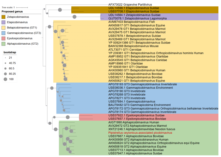
Figure 2. Phylogenetic analysis of RdRP amino acid sequences of Pipistrellus ceylonicus-derived picobirnavirus across the Picobirnaviridae family. The maximum likelihood analysis based on 803 aa alignment was performed using under the LG+I+G substitution model with 1000 bootstrap replicates Colored ranges indicate proposed genera; the sequence titles indicate proposed genus, ICTV-recognized genotype and the isolation source. The newly identified virus is highlighted in red
The Pipistrellus ceylonicus-derived picobirnavirus robustly clusters within ICTV-recognized genogroup 2 and the proposed genus Alphapicobirnavirus. The neighboring clade include sequences identified in samples obtained from human and rodents (Marmota himalayana and Neodon fuscus). The fact that newly observed virus fits into one of the proposed genera additionly supports this proposed division.
The pairwise genetic distances (p-distance) for the capsid protein amino acid sequences are visualized in a distance matrix, which includes the amino acid sequences of the capsid proteins of representative members of Picobirnaviridae family, for which the capsid protein sequence is available in the public databases (Figure 3). The distances for Pipistrellus ceylonicus-derived picobirnavirus are shown in the first column; the titles include the recognized genotypes, the proposed genera titles (for sequences where the corresponding RdRp sequence used for genotyping was available) and the isolation sources of corresponding viruses.
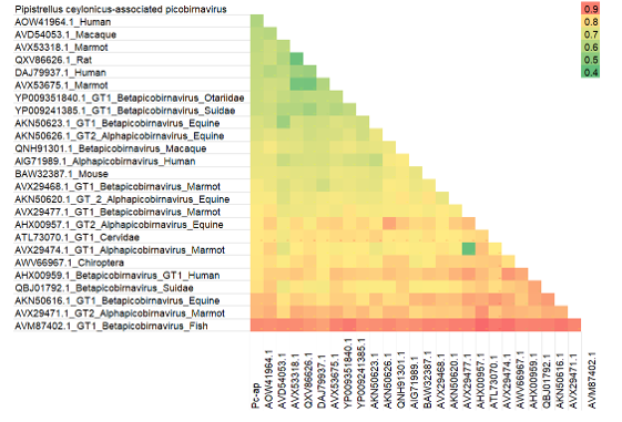
Figure 3. The pairwise distance matrix of the amino acid sequences of the capsid proteins of representative members of Picobirnaviridae family
Notably, sequence diversity among capsid proteins in the Picobirnaviridae family is in general high and shows no correlation with either the RdRP-defined genotype or the isolation host. The Pipistrellus ceylonicus-derived picobirnavirus demonstrates the greatest, yet still low, capsid protein similarity to viruses isolated from a human and a macaque (genetic distances of 0.59 and 0.62, respectively). In contrast, its divergence from AWV66967, the only previously known near-complete picobirnavirus sequence from a chiropteran host, is 0.79, indicating a distant phylogenetic relationship.
Discussion
Significant gaps remain in our knowledge of the Picobirnaviridae family. The absence of a reliable picobirnavirus culture system restricts their detection and characterization to metagenomic sequencing (Kashnikov et al., 2023). As this method has become more accessible, it has facilitated the recent discovery of novel picobirnaviruses in a diverse range of potential hosts, including wild and domestic birds (Pankovics et al., 2018; Ullah et al., 2022), cattle (Huaman et al., 2021; Woo et al., 2019), primates (Woo et al., 2019), marsupials (Chong et al., 2019), pigs (Chauhan et al., 2022), deer (Huaman et al., 2021a, 2021b), bats (Yinda et al., 2018) and various other mammalian species. However, fundamental questions persist regarding its true host range, the relationship between host species and viral genetics, and its potential role in pathogenesis (Kashnikov et al., 2023). The characterization of this novel picobirnavirus contributes to addressing these questions by expanding the known diversity of the family and providing new insights into both its potential host breadth and the composition of the Vietnamese bat virome.
Currently, the Picobirnaviridae family contains only a single recognized genus (Delmas et al., 2019). However, the extreme genetic diversity within this genus (Ghosh et al., 2009) and the broad range of host species from which these viruses have been isolated suggest that the current taxonomy does not accurately reflect their evolutionary relationships. A proposed classification system based on RdRP amino acid sequences defines several distinct genera (Sadiq et al., 2024), which aligns well with the observed clustering of known picobirnavirus sequences. The novel virus described in this study is consistent with this proposed classification system, as it robustly clusters within the proposed Alphapicobirnavirus genus.
Current ICTV species demarcation criteria for Picobirnaviridae are based primarily on host specificity (Delmas et al., 2019). However, the official report notes that, given the substantial genetic distances observed, each picobirnavirus with a complete genome may eventually be classified as a distinct species. The P. ceylonicus-derived picobirnavirus is genetically distant from known bat-associated picobirnaviruses, while its closest relatives were isolated from different hosts (human and macaque), supporting its designation as a novel species within the family. Furthermore, its highest capsid protein similarity is to a human-derived virus rather than to other bat-associated picobirnaviruses.
While a broad correlation exists between genomic lineage and host range for some Picobirnaviridae representatives, such as members of the proposed Gammapicobirnavirus genus (Genotype 3), which are primarily found in environmental and invertebrate samples (Shi et al., 2016), this link is virtually absent among more closely related vertebrate hosts. For example, betapicobirnaviruses have been detected in hosts ranging from fish to humans (Sadiq et al., 2024). The genetic distances between capsid protein sequences, which are theoretically linked to host specificity, show an even weaker association with the isolation source, underscoring the complex and poorly constrained host-virus relationships within this family.
This study has several limitations, partly due to the limited knowledge available regarding the Picobirnaviridae family. Owing to the absence of a culture system, definitive host identification of the newly detected virus is not possible, and in‑depth virological characterization is nearly unattainable. Consequently, as in most studies on Picobirnaviridae, the characterization of novel viral species is based solely on sequences obtained from metagenomic experiments. As previously noted, it remains possible that the virus is associated with the gut bacterial microbiota rather than the bat host itself, or alternatively, that it originates from the bat's diet (e.g., insects). However, the latter scenario appears unlikely, given that the newly identified virus is genetically related to viruses isolated from mammalian feces and distant from known invertebrate‑associated picobirnaviruses. Likewise, the fecal sample collection protocol cannot completely rule out the possibility that the virus originated from the exterior surface of the animal rather than the intestinal tract. Nevertheless, these limitations are typical for animal‑derived viruses that lack established cultivation systems.
Conclusion
While surveillance studies often focus on viral families with established zoonotic and pandemic potential, such as Coronaviridae, Orthomyxoviridae and Flaviviridae, the broader bat virome holds critical insights into the diversity of other understudied viral groups. The Picobirnaviridae family exemplifies this, characterized by a broad host range, substantial genetic diversity, and numerous unresolved questions regarding its ecology and evolution. Answering these questions requires expanded data on both its genetic spectrum and true host associations. The identification of a novel picobirnavirus in P. ceylonicus contributes to this goal by expanding the known host associations and enriching the documented viral diversity within Vietnamese bats.
Funding: This research was supported by the project “Exploring the diversity and circulation of viruses in bat and wild bird populations across tropical habitats” and FMBA of Russia (Registration number 124021900144-4).
Statement on the Use of Generative AI (GenAI): No AI Used
Author Contribution: Conceptualization, Alina Dmitrievna Matsvay; Methodology, Alina Dmitrievna Matsvay and Alexander Pavlovich Yuzefovich; Software, Polina Andreevna Nikolaeva; Validation, Alina Dmitrievna Matsvay; Formal Analysis, Ivan Fedorovich Stetsenko and Polina Andreevna Nikolaeva; Investigation, Ivan Fedorovich Stetsenko, Vasilina Konstantinovna Lapshina, Diana Agaronovna Grigoryan and Alexander Pavlovich Yuzefovich; Resources, Alexander Pavlovich Yuzefovich and Truong Van Tran; Data Curation, Alina Dmitrievna Matsvay; Writing - Original Draft, Ivan Fedorovich Stetsenko; Writing - Review And Editing, Alina Dmitrievna Matsvay and Mo Thi Luong; Supervision, Alina Dmitrievna Matsvay; Project Administration, Truong Van Tran, and Alina Dmitrievna Matsvay; Funding Acquisition, Mo Thi Luong and Alina Dmitrievna Matsvay. All authors have read and agreed to the published version of the manuscript.
Conflict of Interest Statement: The authors declare no conflicts of interest.
References
Altschul, S. F., Gish, W., Miller, W., Myers, E. W., & Lipman, D. J. (1990). Basic local alignment search tool. Journal of Molecular Biology, 215(3), 403–410. https://doi.org/10.1016/S0022-2836(05)80360-2
Arai, S., Aoki, K., Sơn, N. T., Tú, V. T., Kikuchi, F., Kinoshita, G., Fukui, D., Thành, H. T., Gu, S. H., Yoshikawa, Y., Tanaka-Taya, K., Morikawa, S., Yanagihara, R., & Oishi, K. (2019). Đakrông virus, a novel mobatvirus (Hantaviridae) harbored by the Stoliczka's Asian trident bat (Aselliscus stoliczkanus) in Vietnam. Scientific Reports, 9(1), 10239. https://doi.org/10.1038/s41598-019-46697-5
Berg, M. G., Forberg, K., Perez, L. J., Luk, K. C., Meyer, T. V., & Cloherty, G. A. (2021). Emergence of a distinct picobirnavirus genotype circulating in patients hospitalized with acute respiratory illness. Viruses, 13(12), 2534. https://doi.org/10.3390/v13122534
Buchfink, B., Reuter, K., & Drost, H. G. (2021). Sensitive protein alignments at tree-of-life scale using DIAMOND. Nature Methods, 18(4), 366–368. https://doi.org/10.1038/s41592-021-01101-x
Chauhan, R. P., San, J. E., & Gordon, M. L. (2022). Metagenomic analysis of RNA fraction reveals the diversity of swine oral virome on South African backyard swine farms in the uMgungundlovu District of KwaZulu-Natal Province. Pathogens (Basel, Switzerland), 11(8), 927. https://doi.org/10.3390/pathogens11080927
Chong, R., Shi, M., Grueber, C. E., Holmes, E. C., Hogg, C. J., Belov, K., & Barrs, V. R. (2019). Fecal viral diversity of captive and wild tasmanian devils characterized using virion-enriched metagenomics and metatranscriptomics. Journal of Virology, 93(11), e00205-19. https://doi.org/10.1128/JVI.00205-19
Delmas, B., Attoui, H., Ghosh, S., Malik, Y. S., Mundt, E., Vakharia, V. N., & Ictv Report Consortium (2019). ICTV virus taxonomy profile: Picobirnaviridae. The Journal of General Virology, 100(2), 133-134. https://doi.org/10.1099/jgv.0.001186
Ganesh, B., Bányai, K., Kanungo, S., Sur, D., Malik, Y. S., & Kobayashi, N. (2012). Detection and molecular characterization of porcine picobirnavirus in feces of domestic pigs from kolkata, India. Indian Journal Of Virology: An Official Organ of Indian Virological Society, 23(3), 387–391. https://doi.org/10.1007/s13337-012-0106-z
Ghosh, S., Kobayashi, N., Nagashima, S., & Naik, T. N. (2009). Molecular characterization of full-length genomic segment 2 of a bovine picobirnavirus (PBV) strain: evidence for high genetic diversity with genogroup I PBVs. The Journal of General Virology, 90(Pt 10), 2519–2524. https://doi.org/10.1099/vir.0.013987-0
Giordano, M. O., Martinez, L. C., Rinaldi, D., Gúinard, S., Naretto, E., Casero, R., Yacci, M. R., Depetris, A. R., Medeot, S. I., & Nates, S. V. (1998). Detection of picobirnavirus in HIV-infected patients with diarrhea in Argentina. Journal of Acquired Immune Deficiency Syndromes And Human Retrovirology: Official Publication of the International Retrovirology Association, 18(4), 380–383. https://doi.org/10.1097/00042560-199808010-00010
Huaman, J. L., Pacioni, C., Sarker, S., Doyle, M., Forsyth, D. M., Pople, A., Hampton, J. O., Carvalho, T. G., & Helbig, K. J. (2021a). Molecular epidemiology and characterization of picobirnavirus in wild deer and cattle from Australia: Evidence of genogroup I and II in the upper respiratory tract. Viruses, 13(8), 1492. https://doi.org/10.3390/v13081492
Huaman, J. L., Pacioni, C., Sarker, S., Doyle, M., Forsyth, D. M., Pople, A., Carvalho, T. G., & Helbig, K. J. (2021b). Novel picornavirus detected in wild deer: identification, genomic characterisation, and prevalence in Australia. Viruses, 13(12), 2412. https://doi.org/10.3390/v13122412
Inagaki, T., Yamada, S., Fujii, H., Yoshikawa, T., Shibamura, M., Harada, S., Fukushi, S., Le, M. Q., Nguyen, C. T., Nguyen, T. T. T., Nguyen, T. T., Nguyen, T. T., Quach, V. T., Thong, V. D., Mori, K., Sasaki, M., Setiyono, A., Handharyani, E., Takeyama, H., Hasebe, F., … Saijo, M. (2020). Characterization of a novel alphaherpesvirus isolated from the fruit bat Pteropus lylei in Vietnam. Journal of Virology, 94(18), e00673-20. https://doi.org/10.1128/JVI.00673-20
Kashnikov, A. Y., Epifanova, N. V., & Novikova, N. A. (2023). On the nature of picobirnaviruses. Vavilovskii Zhurnal Genetiki i Selektsii, 27(3), 264–275. https://doi.org/10.18699/VJGB-23-32
Kim, B. S., Kim, J. N., & Cerniglia, C. E. (2011). In vitro culture conditions for maintaining a complex population of human gastrointestinal tract microbiota. Journal of Biomedicine & Biotechnology, 2011, 838040. https://doi.org/10.1155/2011/838040
Knox, M. A., Gedye, K. R., & Hayman, D. T. S. (2018). The challenges of analysing highly diverse picobirnavirus sequence data. Viruses, 10(12), 685. https://doi.org/10.3390/v10120685
Koren, S., Walenz, B. P., Berlin, K., Miller, J. R., Bergman, N. H., & Phillippy, A. M. (2017). Canu: scalable and accurate long-read assembly via adaptive k-mer weighting and repeat separation. Genome Research, 27(5), 722–736. https://doi.org/10.1101/gr.215087.116
Kozlov, A. M., Darriba, D., Flouri, T., Morel, B., & Stamatakis, A. (2019). RAxML-NG: a fast, scalable and user-friendly tool for maximum likelihood phylogenetic inference. Bioinformatics (Oxford, England), 35(21), 4453–4455. https://doi.org/10.1093/bioinformatics/btz305
Krishnamurthy, S. R., & Wang, D. (2018). Extensive conservation of prokaryotic ribosomal binding sites in known and novel picobirnaviruses. Virology, 516, 108–114. https://doi.org/10.1016/j.virol.2018.01.006
Kruskop, S. V. (2013). Bats of Vietnam (2nd ed., revised and supplemented; Vol. 1, Biodiversity of Vietnam). Joint Vietnamese- Russian Tropical Science and Technology Research Center & Zoological Museum, Lomonosov Moscow State University.
Lapshina, V. K., Guskova, N. I., Stetsenko, I. F., Luong, M. T., Tran, T. V., Matsvay, A. D., Shipulin, G. A., Yudin, S. M., & Skvortsova, V. I. (2025). Characterizing the bat virome of Vietnam: a systematic review of viral diversity and zoonotic potential. Viruses, 17(12), 1532. https://doi.org/10.3390/v17121532
Latinne, A., Nga, N. T. T., Long, N. V., Ngoc, P. T. B., Thuy, H. B., Predict Consortium, Long, N. V., Long, P. T., Phuong, N. T., Quang, L. T. V., Tung, N., Nam, V. S., Duoc, V. T., Thinh, N. D., Schoepp, R., Ricks, K., Inui, K., Padungtod, P., Johnson, C. K., Mazet, J. A. K., … Fine, A. E. (2023). One health surveillance highlights circulation of viruses with zoonotic potential in bats, pigs, and humans in Viet Nam. Viruses, 15(3), 790. https://doi.org/10.3390/v15030790
Legoff, J., Resche-Rigon, M., Bouquet, J., Robin, M., Naccache, S. N., Mercier-Delarue, S., Federman, S., Samayoa, E., Rousseau, C., Piron, P., Kapel, N., Simon, F., Socié, G., & Chiu, C. Y. (2017). The eukaryotic gut virome in hematopoietic stem cell transplantation: new clues in enteric graft-versus-host disease. Nature Medicine, 23(9), 1080–1085. https://doi.org/10.1038/nm.4380
Li, L., Giannitti, F., Low, J., Keyes, C., Ullmann, L. S., Deng, X., Aleman, M., Pesavento, P. A., Pusterla, N., & Delwart, E. (2015). Exploring the virome of diseased horses. The Journal of General Virology, 96(9), 2721–2733. https://doi.org/10.1099/vir.0.000199
Malik, Y. S., Kumar, N., Sharma, K., Dhama, K., Shabbir, M. Z., Ganesh, B., Kobayashi, N., & Banyai, K. (2014). Epidemiology, phylogeny, and evolution of emerging enteric Picobirnaviruses of animal origin and their relationship to human strains. BioMed Research International, 2014, 780752. https://doi.org/10.1155/2014/780752
McGinnis, S., & Madden, T. L. (2004). BLAST: at the core of a powerful and diverse set of sequence analysis tools. Nucleic Acids Research, 32(Web Server issue), W20–W25. https://doi.org/10.1093/nar/gkh435
Pankovics, P., Boros, Á., Nemes, C., Kapusinszky, B., Delwart, E., & Reuter, G. (2018). Molecular characterization of a novel picobirnavirus in a chicken. Archives of Virology, 163(12), 3455–3458. https://doi.org/10.1007/s00705-018-4012-6
Peng, M.-W., Wu, Y.-H., Ren, Z.-R., Narigen, Hou, R.-S., Xin, G.-Y., Liao, Y.-Q., Wang, J., Le, S.-J., Shi, P.-B., Zhao, H.-L., Deng, Z.-Q., Wang, D.-X., Chaolemen, & Shi, M. (2025). Meta-transcriptomics characterization of individual Marmota sibirica reveals a wide spectrum of viral and bacterial pathogens in Inner Mongolia of China. Microbiology Spectrum, 13(9), e0018025. https://doi.org/10.1128/spectrum.00180-25
Sadiq, S., Holmes, E. C., & Mahar, J. E. (2024). Genomic and phylogenetic features of the Picobirnaviridae suggest microbial rather than animal hosts. Virus Evolution, 10(1), veae033. https://doi.org/10.1093/ve/veae033
Shi, M., Lin, X. D., Tian, J. H., Chen, L. J., Chen, X., Li, C. X., Qin, X. C., Li, J., Cao, J. P., Eden, J. S., Buchmann, J., Wang, W., Xu, J., Holmes, E. C., & Zhang, Y. Z. (2016). Redefining the invertebrate RNA virosphere. Nature, 540(7634), 539–543. https://doi.org/10.1038/nature20167
Tello, M., Oporto, B., Lavín, J. L., Ocejo, M., & Hurtado, A. (2022). Characterization of a carbapenem-resistant Escherichia coli from dairy cattle harbouring blaNDM-1 in an IncC plasmid. The Journal of Antimicrobial Chemotherapy, 77(3), 843–845. https://doi.org/10.1093/jac/dkab455
Ullah, K., Mehmood, A., Chen, X., Dar, M. A., Yang, S., & Zhang, W. (2022). Detection and molecular characterization of picobirnaviruses in the wild birds: Identification of a novel picobirnavirus possessing yeast mitochondrial genetic code. Virus Research, 308, 198624.https://doi.org/10.1016/j.virusres.2021.198624
Walker, B. J., Abeel, T., Shea, T., Priest, M., Abouelliel, A., Sakthikumar, S., Cuomo, C. A., Zeng, Q., Wortman, J., Young, S. K., & Earl, A. M. (2014). Pilon: an integrated tool for comprehensive microbial variant detection and genome assembly improvement. PloS One, 9(11), e112963. https://doi.org/10.1371/journal.pone.0112963
Wick, R., & Volkening, J. (2017). Porechop [Computer software]. GitHub.https://github.com/rrwick/Porechop
Woo, P. C. Y., Teng, J. L. L., Bai, R., Tang, Y., Wong, A. Y. P., Li, K. S. M., Lam, C. S. F., Fan, R. Y. Y., Lau, S. K. P., & Yuen, K. Y. (2019). Novel Picobirnaviruses in respiratory and alimentary tracts of cattle and monkeys with large intra- and inter-host diversity. Viruses, 11(6), 574. https://doi.org/10.3390/v11060574
Wood, D. E., Lu, J., & Langmead, B. (2019). Improved metagenomic analysis with Kraken 2. Genome Biology, 20(1), 257. https://doi.org/10.1186/s13059-019-1891-0
Yan, C., Zhang, C., Xu, L., Meng, F., Wu, J., Tu, C., Li, Y., & He, B. (2019). Metagenomic analysis of bat virome in the Guangxi–Vietnam border area. Scientia Sinica Vitae, 49(3), 266–279. https://doi.org/10.1360/N052018-00191
Yinda, C. K., Ghogomu, S. M., Conceição-Neto, N., Beller, L., Deboutte, W., Vanhulle, E., Maes, P., Van Ranst, M., & Matthijnssens, J. (2018). Cameroonian fruit bats harbor divergent viruses, including rotavirus H, bastroviruses, and picobirnaviruses using an alternative genetic code. Virus Evolution, 4(1), vey008. https://doi.org/10.1093/ve/vey008